storage
Container Selection and Surface Adsorption: Materials, Closures and Peptide Lost to the Wall
Peptide bound to the container wall is routinely mistaken for low potency. Material comparison, closure selection, mitigation options and a procedure for detecting the loss.
Yes — peptides adsorb to the interior surfaces of tubes, vials, tips and plates, and the container material changes how much is lost 1. The mechanism is physical, not chemical: the peptide leaves solution and binds to the wall, so the concentration in the liquid falls while the molecule is unchanged. Nothing in the appearance of the solution reports it, and a nominal concentration calculated from a correctly weighed mass will not report it either.
Mechanism: how peptide leaves solution
Two interactions dominate. Hydrophobic interaction drives non-polar side chains out of water onto any non-polar surface, and the interior of a plastic tube is such a surface. Electrostatic interaction acts between charged residues and charged sites on the material — silanols on glass are ionised at working pH and attract basic residues 1. Hydrogen bonding and van der Waals contact contribute. The result is an equilibrium between peptide in the bulk and peptide bound at the interface.
The same affinity is well described for proteins, where adsorption at solid surfaces and at the air-liquid interface causes not only loss of material but unfolding of the adsorbed layer and aggregation seeded from it 23. Peptides mostly have no tertiary structure to lose, so the usual consequence is plain loss of concentration — but the driving forces are identical.
Two features make this awkward. Adsorption is fast: it can be substantially complete within the time taken to prepare and transfer a dilute solution. And it is not reliably reversible — bound peptide does not necessarily come off when buffer is added, so rinsing the vessel is not a recovery step.
Why concentration decides the size of the loss
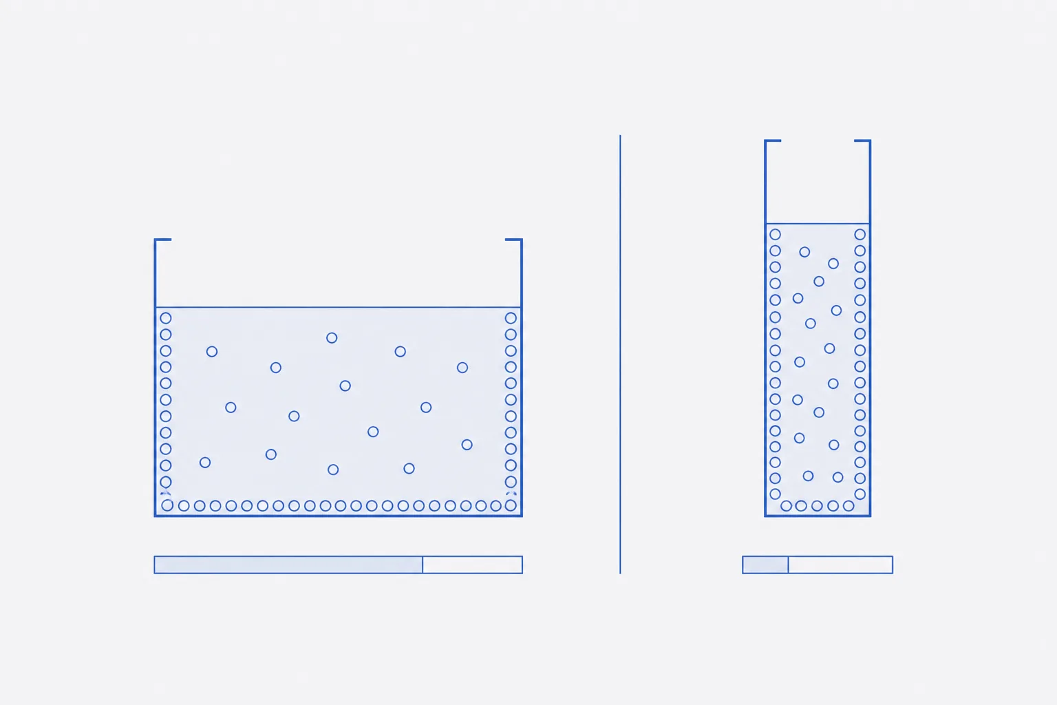
The vessel fixes the surface area. A given tube presents the same wall area to a 100 µM solution as to a 100 nM solution, and the quantity that area accommodates is approximately fixed too, because the surface saturates. What changes with dilution is the amount in the liquid, so the same absolute loss becomes a larger proportion of the total as concentration falls 1.
Hence the working rule. At millimolar and high micromolar concentrations the bound quantity is a negligible fraction of the total. At low micromolar and below it becomes material, and in dilute working solutions a substantial part of the peptide may be on the container rather than in the sample. The magnitude for a given sequence, buffer and vessel must be measured, not assumed 1.
Surface-to-volume ratio is the second lever, and unlike assay concentration it is under the operator's control. A small volume in a large tube loses proportionally more than the same volume in a vessel sized to it. Serial dilution compounds this: each step runs lower than the last, in a fresh vessel with a fresh unsaturated surface.
Container materials compared
The ranking below is general; the order of any two materials can invert for a particular sequence, which is why plasticware and glassware selection for peptide work has been studied as a variable in its own right 1.
| Material | Adsorption tendency | Chemical compatibility | Practical notes |
|---|---|---|---|
| Borosilicate glass | Moderate to high for basic sequences; ionised silanols bind cationic residues | Excellent. Inert to acetonitrile, alcohols, dimethyl sulfoxide, heat and acid | Default for organic-solvent stocks; poor for dilute aqueous basic peptides. Amber grades add light exclusion |
| Silanised glass | Lower than untreated glass; the coating caps silanols and removes the electrostatic contribution | Good, though strong base and prolonged solvent contact degrade the coating | Reproducible while intact. Integrity is not verifiable by inspection — treat as consumable |
| Polypropylene | Lower than plain glass for charged sequences; non-polar, so hydrophobic sequences bind | Good with aqueous buffers and short-chain alcohols; some organic solvents swell it | Usual material for aqueous handling. Surface varies by manufacturer and batch: a change of supplier is a change of variable |
| Low-binding polypropylene | Lowest in routine use. Treated to suppress hydrophobic and ionic binding | As polypropylene; the treatment, not the bulk polymer, limits solvent contact | Correct default below low micromolar. Specify by stated grade — not distinguishable by eye |
| Polystyrene | High. Strongly hydrophobic; untreated tubes and plates bind readily | Poor with organic solvents — crazes in ketones, attacked by dimethyl sulfoxide | Common as untreated plates and disposable tubes, and reached for by default. Avoid for dilute peptide |
| PTFE and fluoropolymer linings | Low to moderate. Non-polar but very low surface energy | Outstanding. Effectively inert to common solvents and extremes of pH | Met as septum facing and cap liners rather than vessel bodies. The choice where solvent contacts the closure |
Sequence and solution factors that raise risk
Material sets the surface. These properties of the peptide and the solution decide how strongly it is engaged, and are worth assessing before a dilute solution is prepared.
- High hydrophobicity. Any sequence needing an organic co-solvent to dissolve is high risk on plastic.
- High net charge. Basic sequences bind ionised silanols on glass; acidic sequences bind amine-functionalised surfaces.
- Small size. Fewer intramolecular contacts compete with the surface, and a given mass is more molecules.
- Low working concentration. The dominant factor, and imposed by the assay rather than chosen.
- Low ionic strength, and pH near the isoelectric point. Both reduce repulsion from a like-charged surface.
- Long holds, many transfers, vigorous mixing. Each transfer presents a fresh unsaturated surface.
Mitigation, and what each measure costs
No mitigation is free. Each works by occupying the surface, weakening the interaction, or raising the concentration — and each puts something into the sample that may itself interfere 2.
| Measure | Mechanism | Limitation |
|---|---|---|
| Carrier protein, typically serum albumin | Saturates surface sites, so the wall holds carrier rather than peptide | Adds a large excess of foreign protein. Rules out mass spectrometry, total-protein assays, and any method where the carrier is detected |
| Non-ionic surfactant at low concentration, polysorbate class | Adsorbs preferentially at solid-liquid and air-liquid interfaces, displacing peptide | Oxidises on storage; degradation products can damage the sample. Interferes with chromatography. Behaviour changes above the critical micelle concentration |
| Organic co-solvent at low percentage | Makes the solvent less polar, lowering the hydrophobic driving force | Restricts container choice; often incompatible with biological assays, and alters the solution chemistry under study |
| Pre-rinsing the vessel with the same peptide solution | Occupies surface sites in advance; the sacrificial rinse takes the loss | Consumes material. Valid only if the rinse is discarded and the vessel not allowed to dry; unusable where it must be quantitatively clean |
| Raising the working concentration | Reduces the bound fraction directly: surface capacity is fixed, the amount in solution is not | Only where the protocol allows. Solubility caps it, and higher concentration raises self-association risk |
| Reducing surface-to-volume ratio: match vessel to volume, cut transfers | Less wall area per unit of solution, fewer fresh unsaturated surfaces | Constrained by consumables and assay format. Shrinks the loss rather than removing it |
The first two are established measures in liquid protein formulation, where interfacial stress is a recognised destabilising factor 24. Moved into an analytical context they carry a cost the formulator does not face: the additive is present when the sample is measured. Choose before the sample is prepared, and record what was added and why.
Closures, septa and extractables
The closure is part of the container system. Sealing performance comes first: a closure that does not seal admits atmospheric moisture to lyophilised material and lets solvent escape from solution. Solvent loss concentrates the sample — an error in the opposite direction to adsorption, which can mask it. A screw cap with a fluoropolymer-faced liner seals more reliably than a push-fit cap.
Second, extractables. Elastomeric septa and cap liners contain plasticisers, curing agents and mould-release residues that migrate into the contents, faster in organic solvent than in aqueous buffer. A fluoropolymer facing on the solution side is the standard control: the sample contacts inert PTFE, the elastomer supplies only mechanical resilience. Puncturing removes that barrier locally.
Third, particulate. Repeated needle penetration of a septum sheds fragments of elastomer into the vessel — a sample-integrity problem and nothing else. Shed particulate raises measured turbidity and can be mistaken for precipitation, nucleates aggregation, and blocks chromatography frits and autosampler lines. Where a vial will be penetrated more than a few times, use a pre-slit septum or split the material across fresh vials.
Headspace, fill volume and labelling
Fill volume sits between two constraints. Too little liquid in too large a vessel raises the surface-to-volume ratio and leaves a large headspace: more wall area per unit of solution, a larger air-liquid interface, more oxygen against methionine, cysteine and tryptophan. Too full leaves no room for expansion on freezing, and a brim-full rigid container can crack or unseat its closure.
The compromise for material that will be frozen is roughly three-quarters of nominal capacity, in a vessel sized to the aliquot rather than to the stock. That sizing limits freeze-thaw cycling and adsorption at once: a fresh unsaturated surface is presented once per aliquot rather than repeatedly to the same material.
Labelling is the part of container selection that fails silently. Adhesive labels lift when frozen and solvent-based ink dissolves on contact with the solvents used to prepare stocks, so both failure modes coincide with the conditions stocks are held under. Use cryogenic-grade labels applied to a clean, dry vessel at ambient temperature, and a solvent-resistant marker. Record identity, concentration, solvent, container grade and date. Label the body, not the cap — a cap moves between vessels.
Procedure: detecting adsorption loss
Adsorption cannot be inferred from appearance, so it must be measured. The comparison below establishes whether the loss is material for a given peptide, solvent, concentration and container. It requires a concentration method — absorbance at 280 nm where tryptophan or tyrosine is present, a colorimetric assay otherwise, or chromatographic peak area against a standard.
- Define the case: peptide, solvent, working concentration, container material and grade, fill volume, hold temperature and duration.
- Prepare a stock high enough that adsorption is negligible — high micromolar or above — and confirm its concentration.
- Dilute to the working concentration directly in the container under test, with at least three replicates per time point.
- Measure immediately after preparation, handling the measurement in vessels of the same material. Record as the value at time zero.
- Hold the remaining replicates undisturbed in the intended container, at the intended temperature, for the interval a real sample would experience.
- Measure again at the end of the hold, by the same method, with the same handling and vessels.
- Run the identical sequence in a low-binding vessel as a comparator, with a solvent blank through both arms.
- Compare. A fall exceeding the repeatability of the method, smaller or absent in the comparator, is adsorption; a fall equally large in both arms is more likely degradation or precipitation.
- Repeat with one mitigation at a time, and record the outcome against the exact combination tested. It does not transfer to another sequence, buffer or concentration.
What makes this worth the effort is the shape of the error. The nominal concentration is calculated correctly, the solution is clear, and the preparation repeats — so the result is reproducible. Only the quantity actually dissolved is wrong, and it is wrong in the same direction every time the same vessel and concentration are used 1. Reproducibility is commonly read as evidence of accuracy; where adsorption is present, the two come apart.