degradation
Oxidation of Susceptible Residues: Vulnerability Order, Mechanism and Control
The detailed treatment of one degradation route. Which residues oxidise and to what, the two mechanistic classes and why they need different controls, and where the oxidant comes from on a laboratory bench.
Methionine is the residue most prone to oxidation, followed by cysteine, then tryptophan, with histidine and tyrosine as secondary targets whose damage usually signals a more aggressive oxidant or a bound transition metal 12. That ranking decides whether a preparation needs oxidative controls at all. What follows is the detailed treatment of one route: products and mass changes residue by residue, the two mechanistic classes and why they answer to different controls, the oxidant sources on an ordinary bench, the analytical signature, and a procedure.
Residues in order of vulnerability
Methionine leads because its thioether sulfur is an accessible nucleophile needing no catalyst beyond dissolved oxygen and time. The first product is the sulfoxide, one oxygen added, plus 16 Da; a stronger oxidant takes the sulfoxide sulfur to the sulfone, plus 32 Da from the parent 1. The two differ in more than mass — the sulfoxide can be chemically reduced back, the sulfone cannot. A short synthetic sequence has no folded core to bury a methionine in, so intrinsic reactivity sets the rate.
Cysteine is second and has more destinations. The reactive species is the thiolate rather than the thiol, so reactivity climbs as pH rises through the sulfhydryl dissociation region. The mildest outcome is pairing to a disulfide, minus 2 Da: intended where a bridge is designed, unintended where it is not, giving dimers and mispaired isomers. Beyond that the sulfur climbs an oxygen ladder — sulfenic acid at plus 16 Da, unstable; sulfinic at plus 32 Da; sulfonic at plus 48 Da, terminal 13.
Tryptophan is third and the most varied. The indole ring gives hydroxytryptophan at plus 16 Da, ring-opened N-formylkynurenine at plus 32 Da, and kynurenine at plus 4 Da once the formyl group is lost from N-formylkynurenine 1. A tryptophan-containing preparation therefore presents not as one new peak but a family of them. The intact indole carries the residue's fluorescence, so loss of it screens for damage at no instrument cost.
Histidine and tyrosine are secondary. Histidine gives 2-oxohistidine at plus 16 Da, and matters out of proportion to its reactivity because the imidazole nitrogen is a common transition-metal ligand: the histidine is often both anchor for the metal and first casualty of what follows 1. Tyrosine gives the catechol 3,4-dihydroxyphenylalanine at plus 16 Da, and phenoxyl radicals on two tyrosines couple to a dityrosine crosslink — a dimer at twice the monomer mass minus 2 Da, leaving monomer mass unchanged and so invisible to intact-mass measurement 2.
| Residue | Oxidation product | Mass change | Note |
|---|---|---|---|
| Methionine | Methionine sulfoxide | +16 Da | Usual first observation; chemically reversible |
| Methionine | Methionine sulfone | +32 Da from parent | Forcing conditions; irreversible |
| Cysteine | Disulfide, intra- or intermolecular | −2 Da per pair | Intended where a bridge is designed |
| Cysteine | Sulfenic acid | +16 Da | Unstable; reacts onward |
| Cysteine | Sulfinic acid | +32 Da from parent | Effectively irreversible |
| Cysteine | Sulfonic acid | +48 Da from parent | Terminal state |
| Tryptophan | Hydroxytryptophan | +16 Da | First of a family |
| Tryptophan | N-formylkynurenine | +32 Da | Ring opened; fluorescence lost |
| Tryptophan | Kynurenine | +4 Da from parent | Formyl lost from the preceding product |
| Histidine | 2-oxohistidine | +16 Da | Frequent metal ligand |
| Tyrosine | 3,4-dihydroxyphenylalanine | +16 Da | Catechol product |
| Tyrosine | Dityrosine crosslink | Dimer at 2M − 2 Da | Monomer mass unchanged |
Read the sequence first
A sequence containing no methionine, cysteine, tryptophan, histidine or tyrosine is not at risk from this route at all. No chelator, inert overlay or amber vial improves it, because nothing is present for an oxidant to attack. Worth stating plainly: oxidative controls are often applied as general precaution to material that cannot benefit, while the routes that will actually consume it go unaddressed.
So the first step is arithmetic on the sequence, not a decision about storage. Count the susceptible residues; note whether any cysteine is free or paired as designed; note whether the sequence presents a plausible metal-binding arrangement — more than one histidine, adjacent aspartate or glutamate carboxylates, or a free N-terminal amine beside a histidine. Those three observations determine which controls below are worth the trouble.
Two mechanistic classes
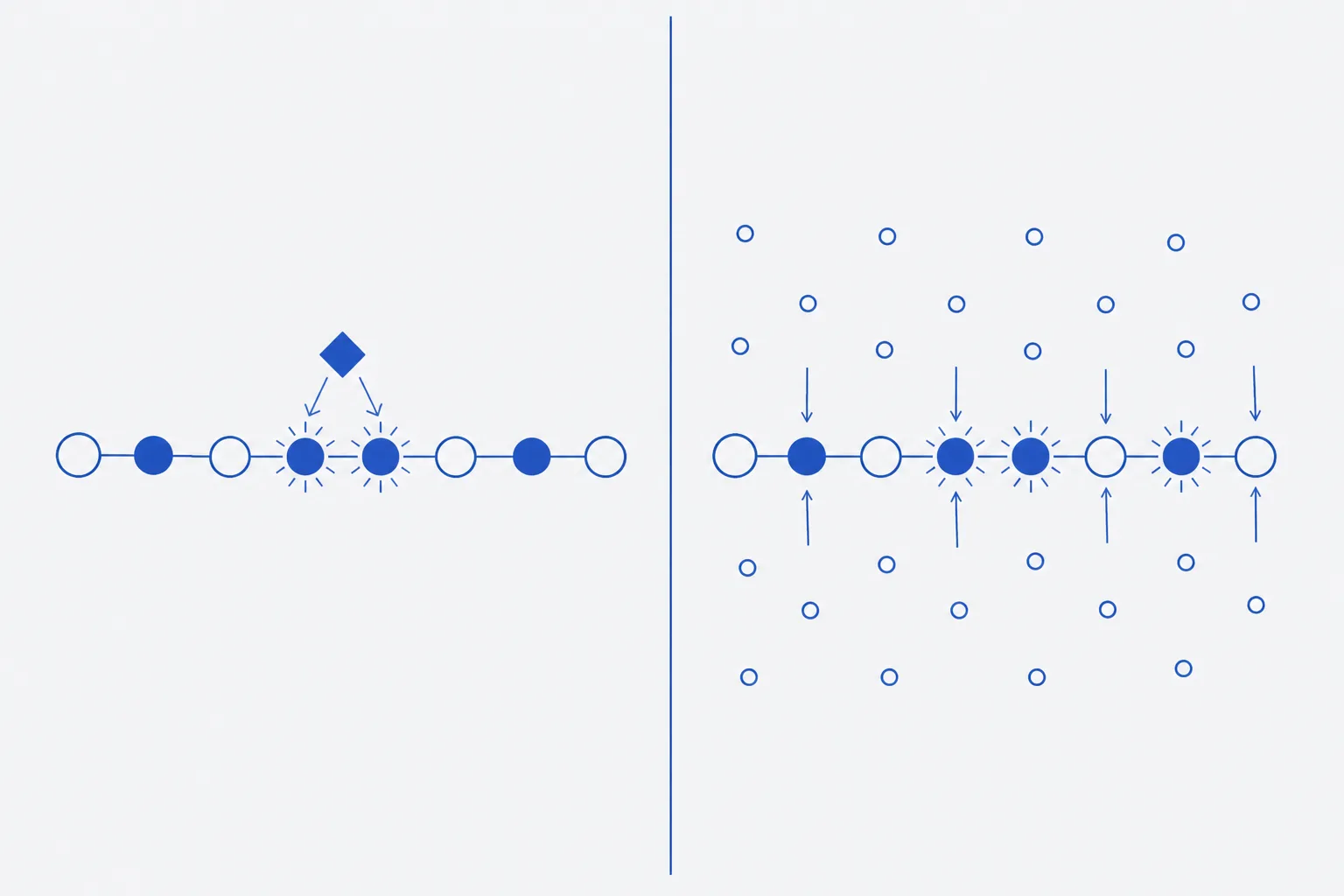
Oxidative damage arrives by two mechanisms usually described together, which they should not be. In site-specific metal-catalysed oxidation, a transition metal ion — iron or copper — binds at a site on the peptide, is reduced by whatever ambient reductant is present, and reacts with oxygen or hydrogen peroxide to generate a reactive oxidant in place. Because that oxidant is produced on the binding site rather than delivered to it, it reacts with whatever is adjacent before it can diffuse. Damage is localised: residues near the metal site are attacked while identical residues elsewhere in the same molecule are untouched 1.
Three consequences follow. Trace metal levels well below anything registering as contamination suffice, because the metal turns over catalytically rather than being consumed. The reaction is not photochemical, so amber glass, foil and dark storage do nothing whatever to prevent it. And the countermeasure is not oxidant exclusion but chelation, which removes the metal from the peptide site rather than the oxidant from the solution 13.
General oxidation is the other class: dissolved molecular oxygen, hydroperoxides carried in by a solvent or excipient, or reactive species generated by light acting on a sensitiser. The oxidant is present throughout the bulk and arrives by diffusion, so damage distributes in rough proportion to intrinsic reactivity rather than concentrating at one point in the chain 2. Every copy of a susceptible residue meets the same insult. This class does answer to oxidant exclusion, inert headspace, sacrificial scavengers and, where light drives it, opaque containment.
The distinction is diagnostic as well as mechanistic. A map showing one methionine heavily oxidised while a second in the same sequence is untouched points to the site-specific mechanism; comparable oxidation at both points to a bulk oxidant. The inference is suggestive rather than conclusive, since sequence context also modulates reactivity, but it decides whether to reach for a chelator or a nitrogen line.
Sources of oxidant on a laboratory bench
The reagents for this route are ambient, which is why it is met so often, and identifiable objects rather than abstractions, which is what makes it controllable.
- Peroxide accumulation in aged solvents. Diethyl ether, di-isopropyl ether, tetrahydrofuran and dioxane autoxidise on standing in contact with air, accumulating hydroperoxides, and light accelerates it. Ether is in routine use for precipitating crude peptide after cleavage, so an old bottle is often the largest single oxidant source in a peptide laboratory. Date bottles on opening; test with peroxide strips before use.
- DMSO. An oxidant, not an inert solvent. It converts thiols to disulfides — a reaction used deliberately to close bridges — and thioethers to sulfoxides, faster when warm or acidic. A DMSO stock of a cysteine- or methionine-containing sequence is an oxidation experiment nobody meant to set up.
- Trace iron and copper. Ordinary-grade buffer salts, glass surfaces, stainless steel in contact with solution and water below its stated specification introduce transition metals far beneath visibility and far above what site-specific catalysis needs.
- Headspace oxygen. Air above the liquid in a partly used vial is a reservoir replenished at every opening, and buffer equilibrated with air carries dissolved oxygen at the low milligram-per-litre level. Neither is dramatic; both act for the whole storage interval.
- Light. Laboratory lighting and daylight drive photo-oxidation and generate reactive species from sensitisers in solution, tryptophan and tyrosine being both targets and participants.
Analytical signature
Oxidation is among the more tractable routes to identify, because added oxygen is visible as mass. An intact deconvoluted spectrum measured against a fresh reference shows the plus 16, plus 32 and plus 48 series directly, and mapping the digested material localises a shift to a named residue — which converts an ambiguous plus 16 Da into a definite methionine sulfoxide 4. The plus 4 Da of kynurenine and the minus 2 Da of disulfide formation are easier to overlook, but neither hides inside the isotope envelope as a single-dalton shift does.
Two confounders. Oxidation can be introduced during sample preparation and in the ion source, so an apparent plus 16 Da is a claim about the sample as measured, not as stored; a fresh reference through the identical workflow on the same day is the control. And the dityrosine crosslink changes no monomer mass, so an intact-mass result finding nothing does not exclude oxidative damage where tyrosine is present 2.
Chromatography supplies the second signature. Adding oxygen to a thioether or an indole raises polarity, so oxidised species are retained less strongly on reverse phase and a methionine sulfoxide typically elutes earlier than its parent under the same gradient. The characteristic presentation is a new shoulder or resolved peak ahead of the main peak carrying a plus 16 Da mass — so inspect that region first. Loss of tryptophan fluorescence screens for indole damage, and a free-thiol assay reports cysteine pairing that chromatography may not resolve.
Control measures and their limitations
Each control addresses one mechanism and leaves the other largely intact, so the choice follows the mechanism identified rather than a general wish to be careful 13.
| Control | Mechanism addressed | Limitation |
|---|---|---|
| Chelator at a defined metal ratio | Metal-catalysed oxidation; strips the metal from the peptide site | No effect on oxygen or peroxide already present; a poor ratio can turn pro-oxidant |
| High-purity salts, specified water, minimal metal contact | Metal input at source, upstream of the same mechanism | Cannot remove metal already carried in with the material |
| Inert headspace, nitrogen or argon; filled vials | General oxidation by dissolved and headspace oxygen | Lost at every opening; no effect on metal-catalysed chemistry |
| Aliquoting into single-use portions | Repeated headspace replacement across the stock | Costs one careful session; no help once oxidant is present |
| Sacrificial free methionine | General oxidation; consumes bulk oxidant first | Consumed over time; adds a component to every analysis; poor cover at a metal site |
| Avoiding DMSO for cysteine and methionine sequences | Removes an oxidant mistaken for an inert solvent | Some sequences need it for solubility; then fresh bottle, cold, short interval |
| Fresh, peroxide-tested solvent; dated bottles | Peroxide-driven oxidation of methionine and tryptophan | Strips detect hydroperoxide, not every oxidant |
| Amber or opaque containment | Photo-oxidation only | No effect on metal-catalysed or dark autoxidation |
| Cold, dry, lyophilised holding | Slows every route including this one | Oxidation continues in the solid state; slower is not stopped |
Handling an oxidation-prone sequence
- Read the sequence and count the methionine, cysteine, tryptophan, histidine and tyrosine residues. If there are none, stop: this route does not apply.
- Record whether any cysteine is free or already paired as designed, and whether the sequence presents a plausible metal-binding arrangement.
- Choose the solvent on that basis. Avoid DMSO where cysteine or methionine is present; where solubility forces it, use a fresh dry bottle, keep the solution cold and the interval short.
- Test any ethereal solvent for peroxide before use. Discard undated bottles rather than testing them.
- Prepare buffers from high-purity salts and water of stated specification. Add a chelator where a metal-binding arrangement is present.
- Aliquot into single-use portions at first opening, and overlay with nitrogen or argon where methionine, cysteine or tryptophan is present.
- Hold in amber or opaque containment, and do not let that stand in for chelation or oxygen exclusion.
- Record a baseline before storage: intact mass, and a reverse-phase profile on the column and gradient to be used later.
- On re-analysis, inject the aged aliquot and a fresh reference on the same day, same column, same workflow. Inspect the region ahead of the main peak first.
- Where a plus 16 Da species is found, map it before assigning it. Localisation separates a site-specific pattern from a bulk oxidant, and so names the control to change.
- Change one condition, re-test a fresh aliquot, and document the previous condition.
Oxidation is the most controllable of the chemical degradation routes precisely because its reagents are objects rather than conditions: a bottle of ether, a grade of buffer salt, the air above a liquid, a lamp above a bench. The corresponding error is to treat the class as one problem with one countermeasure. An amber vial holding a peptide with a copper ion bound to its histidines is not protected; it is only dark 1.