D-Amino Acid Content of Peptides: Marfey's Reagent Procedure
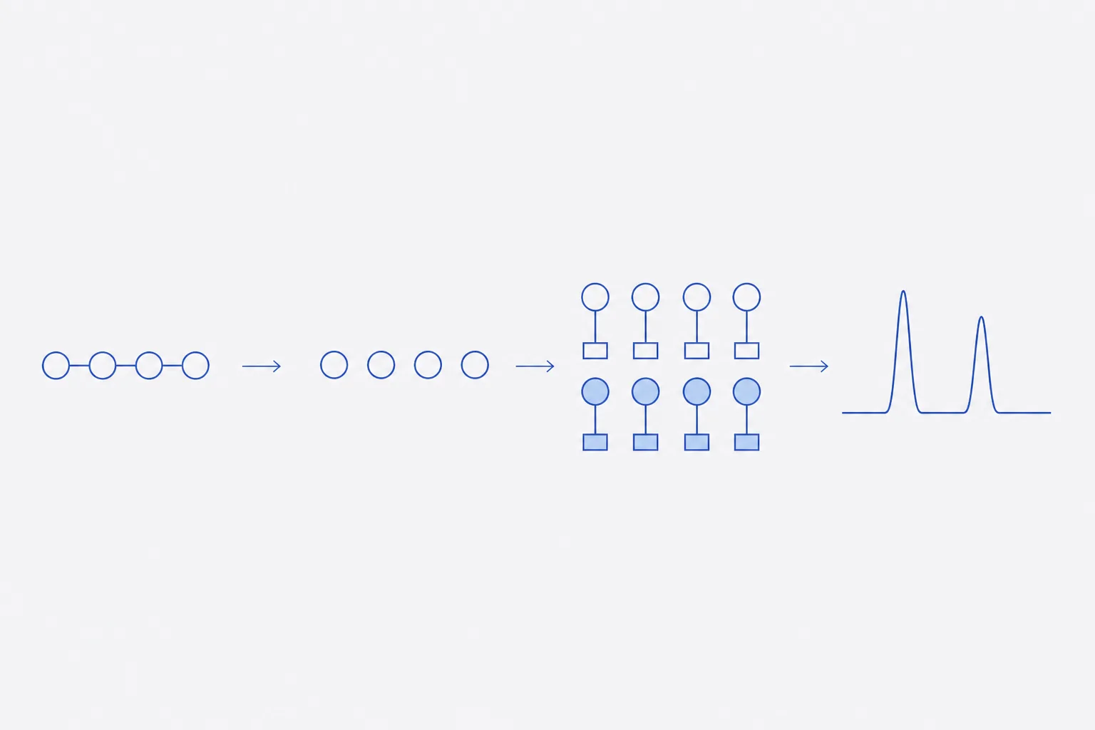
How the D-residue content of a synthetic peptide is measured: hydrolysis in deuterated acid to tag racemisation caused by the method itself, derivatisation with Marfey's reagent, achiral LC-MS separation of the diastereomers, and the per-residue % D calculation with its controls.
The D-residue content of a synthetic peptide is measured by hydrolysing it to free amino acids, converting each amino acid to a diastereomer with a chiral derivatising reagent such as Marfey's reagent, separating the diastereomers on an ordinary reversed-phase column and calculating the percentage of the D form for each residue 13. Hydrolysis itself racemises a small fraction of every residue and would be counted as sample. Carrying out the hydrolysis in deuterated acid labels each residue that inverts during the method with a deuterium atom, a mass increase of about 1 Da, so inversion produced by the method is separated from the D-content already present 1.
The companion reference on synthesis-related impurities names racemisation as a class and gives no measurement. This page gives the measurement as a numbered procedure, the elution-order and control tables, and a worked calculation for one residue of an invented peptide. Typical reagent amounts and times are those reported in the cited method literature; they are starting points to confirm on the laboratory's own standards, not validated settings.
Where D-residues come from
A peptide assembled from L-amino acids should contain only L-residues. D-forms enter in four ways. The protected amino acid used for coupling may contain a trace of its enantiomer. Activation of the carboxyl group for coupling can epimerise the residue being added, most readily for cysteine and histidine. Basic conditions in deprotection can epimerise residues, and aspartimide formation, a ring closure at aspartic acid, can reopen to give D-aspartyl products. Last, storage in solution at high pH can invert residues slowly. Each route produces a diastereomer of the target: same sequence, same mass, different configuration at one position.
Such an impurity is a problem for a reference-method reason. It has the same mass as the target, so mass spectrometry of the intact peptide cannot see it, and it can elute close to the main peak in RP-HPLC or co-elute entirely. Amino acid analysis by ion-exchange or achiral derivatisation also does not distinguish the enantiomers. The configuration of each residue is only visible after the chain is broken down and the enantiomers are resolved.
Why total hydrolysis is needed, and why it racemises
To measure every residue, every peptide bond has to be cleaved. The standard route is 6 mol·L⁻¹ hydrochloric acid at 110 °C for 24 hours in a sealed, evacuated tube. Under those conditions the alpha-hydrogen of an amino acid can exchange with the solvent, and exchange followed by reprotonation can produce either configuration. The inversion is small for most residues and greater for a few, notably Asp, Ser and Cys, and its size depends on the peptide sequence and the hydrolysis time 13.
The consequence is that a sample hydrolysed in ordinary acid cannot report a D-content below the racemisation of the hydrolysis itself, however sensitive the chromatography. Shorter or milder hydrolysis reduces the effect but not to zero and risks incomplete cleavage of hindered bonds, such as those between hydrophobic residues.
Deuterated acid hydrolysis and the +1 Da label
Goodlett and co-workers replaced HCl and water with DCl and D₂O 1. When a residue's alpha-hydrogen is exchanged during hydrolysis, it is replaced by deuterium, and the residue carries a mass one unit heavier than normal. A residue that was already D in the sample, and which did not exchange, has the ordinary mass. The method then resolves four species for each amino acid, as the table shows.
| Species | Mass relative to normal | Origin | Counts toward sample % D? |
|---|---|---|---|
| L, unlabelled | 0 | L-residue in the sample, unchanged | Denominator |
| D, unlabelled | 0 | D-residue already present in the sample | Numerator |
| L, +1 Da | +1.006 Da | Exchanged during hydrolysis; may have come from an L or a D residue | No: method-induced |
| D, +1 Da | +1.006 Da | Exchanged during hydrolysis; may have come from an L or a D residue | No: method-induced |
An exchanged residue forgets its original configuration, so its D or L designation says nothing about the sample. A residue that did not exchange keeps both its mass and its configuration. Only the unlabelled pair therefore reports the sample, which is what the formula uses. One practical correction applies: the carbon-13 isotope peak of an unlabelled residue falls at the same nominal mass as the +1 Da species, so the +1 Da channel needs the natural isotope contribution of the unlabelled peak subtracted. The correction is not needed for the unlabelled channel.
Derivatisation with FDAA
Marfey's reagent is 1-fluoro-2,4-dinitrophenyl-5-L-alanine amide (FDAA). It reacts through its fluorine with the free amino group of an amino acid under mild base, and the product carries a dinitrophenyl chromophore that absorbs strongly near 340 nm 23. A chiral reagent and a chiral amino acid give diastereomers whose physical properties differ, so L-amino acid and D-amino acid derivatives can be separated on an achiral column. Fujii and co-workers combined the method with mass spectrometry and showed how the reagent assigns absolute configuration for each residue in a peptide, using the related leucine-amide reagent 4.
- Hydrolyse the peptide, typically 0.1 to 1 mg, in 6 mol·L⁻¹ DCl in D₂O at 110 °C for 24 hours in a sealed, evacuated or argon-flushed tube. Hydrolyse an all-L reference peptide and a blank tube in the same run.
- Evaporate the acid under vacuum or a stream of nitrogen without heating above room temperature. Redissolve the residue in a small volume of water, so that excess acid does not neutralise the base in the next step.
- Add aqueous sodium hydrogen carbonate, about 1 mol·L⁻¹, to give a mildly basic solution. Add a solution of FDAA, about 1% w/v in acetone, in at least a two-fold molar excess over the total amino groups.
- Mix and heat at about 40 °C for about 1 hour, with the tube protected from light. Derivatisation is complete only if every residue has reacted; excess reagent is required.
- Quench by cooling and adding hydrochloric acid, about 2 mol·L⁻¹, in a volume that neutralises the base. The pH should be near neutral or slightly acid so that the derivatives are stable.
- Dilute with the starting mobile phase, filter, and inject without delay or store at low temperature. State the maximum hold time in the procedure.
Separation and elution order
A C18 column with a water and acetonitrile gradient and an acidic modifier separates the derivatives. Detection is by absorbance at 340 nm, where the reagent chromophore absorbs and most peptide-related interferences do not, and by mass spectrometry for the labelled species. For most residues the L-L diastereomer elutes before the L-D diastereomer, because intramolecular hydrogen bonding makes the L-D form more hydrophobic 35. The order is a rule of thumb. Assign each residue against derivatised pure D- and L-standards run under the same conditions rather than trusting the order.
| Item | Rule or control | Acceptance check |
|---|---|---|
| Elution order | L-L before L-D for most amino acids; exceptions exist, for instance residues with more than one reactive group | Assign every residue from standards, not from the rule |
| Resolution | Separate the D and L derivative of each residue | Resolution of at least 1.5 on the standard mixture |
| Reagent purity | Derivatise an authentic L-amino acid with FDAA and look for the D-derived peak | Impurity from the reagent below the reporting limit |
| Reference peptide | Hydrolyse an all-L peptide of similar composition in DCl/D₂O in the same run | Unlabelled D below the reporting limit; the +1 Da pair reports the method racemisation |
| Hydrolysis symmetry | The +1 Da D and +1 Da L from an all-L peptide should both appear | Method-induced inversion measured and recorded per residue |
| Reagent blank | Reagents and tube without peptide | No peaks at the retention times of the residues |
| Mass accuracy | Extracted ion for each derivatised residue | Within the instrument's stated mass tolerance |
| Completeness | Excess reagent present after the reaction | A reagent peak remains in the chromatogram |
LC-MS detection and assignment
Mass spectrometry separates the unlabelled and the +1 Da species that UV cannot tell apart. Extract the ion for each derivatised amino acid, the mass of the residue plus the reagent minus the leaving group, at its unlabelled mass and at +1.006 Da, and integrate each over the same retention window as the standards. Accurate-mass instruments give a narrow extraction window and so reduce interferences from the peptide matrix 14. The assignment of the D peak in the sample is made on retention time against the derivatised standard, the mass and the isotope pattern together; all three must agree.
Calculating % D per residue and the detection limit
For each residue, take the integrated area of the unlabelled D peak and the unlabelled L peak and calculate % D = D ÷ (D + L) × 100. The worked example is the alanine residue of an invented peptide, hydrolysed in DCl/D₂O and derivatised with FDAA. Areas are extracted-ion areas in arbitrary units, corrected for the carbon-13 contribution in the +1 Da channel.
| Species | Area | Use |
|---|---|---|
| L-Ala, unlabelled | 1,842,000 | Denominator |
| D-Ala, unlabelled | 3,870 | Numerator |
| L-Ala, +1 Da | 9,100 | Method-induced |
| D-Ala, +1 Da | 9,400 | Method-induced |
% D in the sample = 3,870 ÷ (3,870 + 1,842,000) × 100 = 0.21%. Method-induced inversion = (9,100 + 9,400) ÷ (1,842,000 + 3,870 + 9,100 + 9,400) × 100 = 0.99%. Had the sample been hydrolysed in ordinary acid, the D peak would have been the sum of the original and the induced D, 3,870 + 9,400 = 13,270, giving 0.71% D, which is about 3.4 times the true value. The deuterium label is what recovers the difference.
The detection and quantitation limits are those of the minor D peak beside the large L peak, not of the derivative alone. Determine them by spiking authentic D-amino acid into the L-form at a series of fractions, such as 0.05, 0.1, 0.2, 0.5 and 1%, deriving as for samples and finding the lowest fraction at which the D peak is detected with a signal-to-noise of 3 and quantified with acceptable precision at a signal-to-noise of 10. Report results below the quantitation limit as below the stated limit, together with the mass analysed.
Limits of the method and the record
The method reports an average over all copies of each residue type. If the sequence contains three alanines and one is partly D, the result is the % D of the pooled alanine, not of any one position. It cannot say which position is affected: that needs peptide mapping with a chiral or ion-mobility separation, or synthesis of the candidate diastereomer as a reference. Tryptophan is destroyed by acid hydrolysis; asparagine and glutamine are converted to aspartic and glutamic acid and are reported together with them. Cysteine and methionine need protection or oxidation before hydrolysis for accurate recovery.
- Record the peptide batch, the mass hydrolysed with its balance printout, and the DCl/D₂O lot.
- Record hydrolysis time, temperature and tube atmosphere, with the all-L reference run alongside.
- Record the reagent lot and preparation date, the derivatisation time and temperature, and the quench.
- Keep the chromatograms for standards, controls, blanks and samples with the extracted-ion windows used.
- Report % D per residue, the method-induced inversion per residue, the residues not assessed (Gly, Trp and others) and the detection limit for the run.
- Retain data files and audit trail with the report, with reasons for any reintegration.
References
- Peptide chiral purity determination: hydrolysis in deuterated acid, derivatization with Marfey's reagent and analysis using high-performance liquid chromatography-electrospray ionization-mass spectrometry
- Determination of D-amino acids. II. Use of a bifunctional reagent, 1,5-difluoro-2,4-dinitrobenzene
- Marfey's reagent for chiral amino acid analysis: a review
- A Nonempirical Method Using LC/MS for Determination of the Absolute Configuration of Constituent Amino Acids in a Peptide: Combination of Marfey's Method with Mass Spectrometry and Its Practical Application
- Use of Marfey's reagent and analogs for chiral amino acid analysis: assessment and applications to natural products and biological systems