Agitation and Interfacial Stress: Shaking, Vortexing, Sonication
Why mechanical handling damages peptides in solution through interfaces rather than through the bulk liquid, how shaking, vortexing and sonication differ, which variables set the exposure, and a numbered shaking-stress study with unstressed and surfactant controls read by turbidity, size-exclusion chromatography and thioflavin T.
Shaking, vortexing and sonication damage peptides in solution mainly by creating and renewing interfaces: air-water boundaries, bubble walls, foam and container surfaces, where molecules adsorb, partly unfold and associate, rather than by any force acting on the bulk liquid. Reviews of protein aggregation list agitation among the principal induction factors and identify the air-liquid interface as a central route 1. The size of the effect depends on the interfacial area the handling generates, the headspace, the container and the sequence, so a bench procedure that controls those variables controls most of the risk. A solution that looks clear after agitation is not evidence that nothing has happened.
The companion references treat aggregation mechanisms and dissolution. This page isolates the mechanical driver and gives a study design for testing it. Much of the quantitative literature concerns larger proteins, antibodies in particular; where a statement rests on that work, the text says so. Figures in the examples are illustrative and describe no real material.
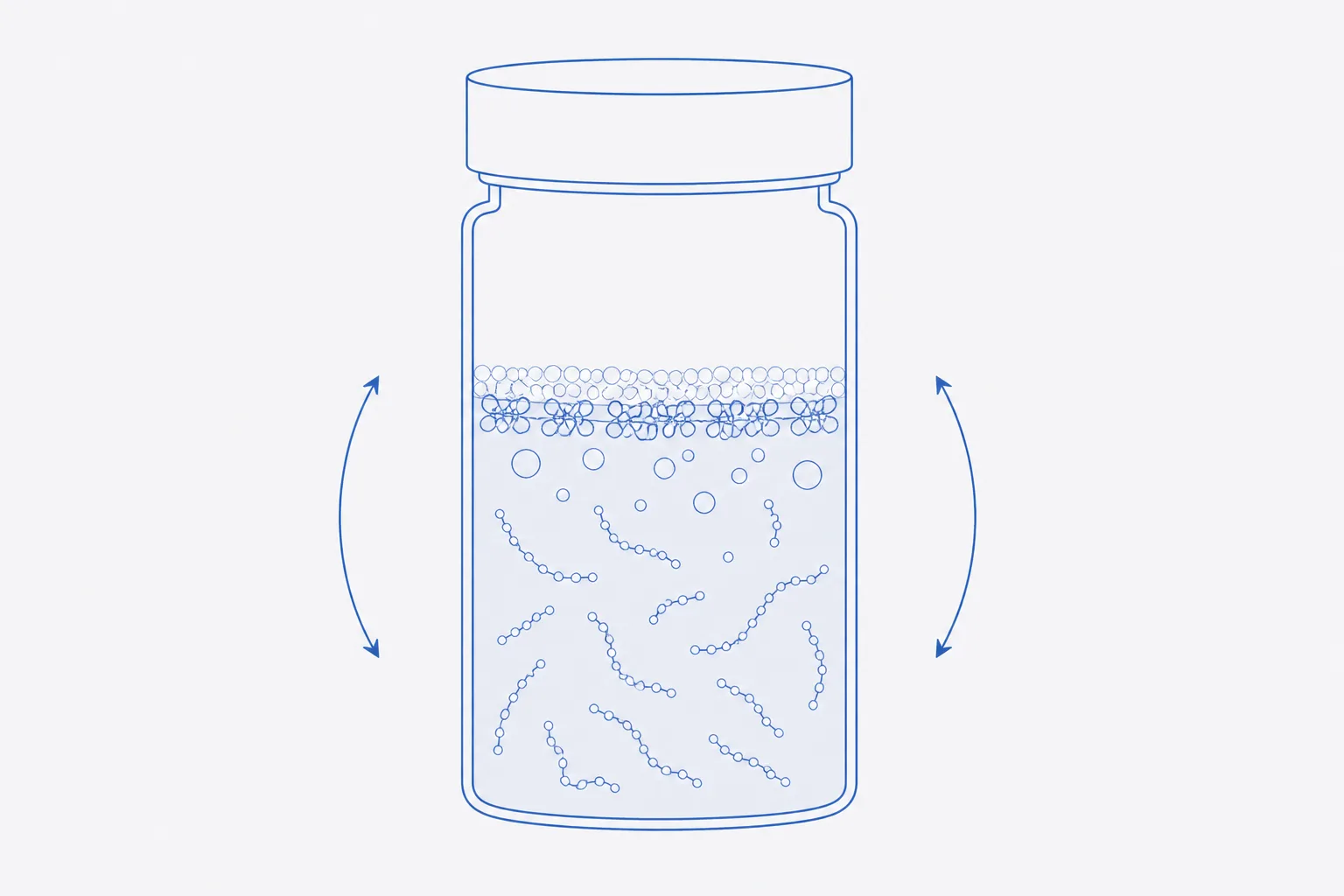
Why interfaces, not bulk solution, drive mechanical aggregation
A peptide with hydrophobic residues lowers its free energy by moving from water to a boundary where those residues can face air or a hydrophobic surface. At the boundary the local concentration rises, the chain can adopt conformations not available in solution and neighbouring molecules can associate. Bulk liquid, by contrast, exerts little force on a small peptide: flow shears that are damaging to long, extended proteins are modest at the length scale of a peptide of a dozen residues. Consistent with this, mechanical-stress studies of an antibody found that conditions which increase the air-liquid interface produce much more aggregation than conditions which stir without entraining air 2.
Agitation matters because it renews the boundary. A quiescent solution reaches an adsorbed layer that then shields the interface; shaking breaks that layer, folds it into the bulk as aggregate and exposes fresh surface for the next cycle. The adsorbed material is partly returned to solution in a changed state, which is why the damage accumulates with time and number of cycles rather than saturating.
The tier of evidence matters. The mechanisms above are established for proteins, particularly antibodies, in vitro 12. For short peptides the same direction of effect is expected, and sequences rich in hydrophobic or aromatic residues are the more susceptible, but the magnitude varies with sequence and has to be measured for the material in hand.
Shaking and vortexing: foam, bubbles and surface renewal
Interfacial area is simple geometry before agitation and unbounded after it. A vial with an inner diameter of 11 mm has a flat liquid surface of about 0.95 cm². One millilitre of solution in it presents roughly 0.95 cm² of air-water interface per millilitre. Vortexing for a few seconds can disperse the headspace as bubbles of 0.1 to 1 mm diameter, and each bubble is a sphere of interface; a foam layer a few millimetres deep can carry an area one to two orders of magnitude larger than the flat surface. These are order-of-magnitude figures, not a prediction for any vial, but they show why the same solution can survive a week undisturbed and fail within a minute of vortexing.
Shaking on an orbital or reciprocating platform generates less foam than a vortex mixer but works for longer. In the antibody work, the extent of aggregation under shaking depended strongly on the headspace and on the presence of a surfactant, which shows that the interface, not the motion itself, was the active variable 2. Rocking in a completely full vial, with no headspace, removes the air-water interface and the effect with it, though not the container wall.
Sonication: cavitation, local heating and radical chemistry
Sonication adds three mechanisms to the interfacial one. Cavitation bubbles grow and collapse thousands of times a second, creating new interface at every cycle. The collapse produces transient local temperatures far above the bulk reading, so material at the bubble wall is heated even if the thermometer is not. And in water, collapse can generate hydroxyl and hydrogen radicals, which oxidise susceptible side chains. Sonication of proteins has been reported to produce aggregates that resemble amyloid, with the aggregation increasing with sonication time 3.
For a peptide, the chemical consequences matter as well as the physical ones. Methionine, cysteine, tryptophan, tyrosine and histidine are the residues most exposed to radical attack, and the oxidation products are unavoidably different species that no later gentler handling will reverse. A bath sonicator is milder than a probe; a short, cooled burst is milder than a continuous one; and sonication used to speed dissolution is a rescue for a solubility problem that should be solved with the solvent instead.
| Operation | Interface generated | Additional stress | Relative risk |
|---|---|---|---|
| Probe sonication | Very large: cavitation bubbles renewed continuously | Local heating; radicals; probe-tip metal release | Highest |
| Bath sonication | Large: cavitation, less intense | Bath heating; radicals at lower rate | High |
| Vortex mixing | Large: foam and entrained bubbles | Surface shear at the vial wall | High |
| Vigorous manual shaking | Moderate to large: foam on each stroke | Warming from the hand | Moderate to high |
| Orbital or reciprocating shaking | Moderate: continuous surface renewal, little foam at low speed | Long duration | Moderate |
| Repeated pipetting with bubbles | Small to moderate: bubble entrainment, wall contact | Tip-surface adsorption | Low to moderate |
| Gentle inversion or swirling | Small: flat surface only | None significant | Low |
| Standing, no headspace | Container wall only | None | Lowest |
Headspace, fill volume and container material as variables
Three variables set the exposure for a given motion. Headspace volume determines how much air is available to be entrained; a half-filled vial agitates far more air than a nearly full one. Fill volume fixes the ratio of interface to liquid, so that the same area is spread over a smaller amount of peptide in a small fill, and the fraction lost rises. And container material decides what the wall contributes: surfaces and leachables are recognised sources of instability for biopharmaceuticals, and hydrophobic polymer walls and silicone oil in particular can promote adsorption and aggregation 4. The companion reference on container selection covers the wall in detail.
Designing a shaking-stress study with proper controls
A shaking-stress study asks whether a preparation tolerates the handling it will receive. The design below is generic; the values in brackets are examples to be replaced by those the question requires, and every one must be recorded.
- Define the question: which handling is being tested (transport, mixing, reconstitution) and which read-out would show failure. Write the acceptance criterion before the run, for example no change in recovered monomer beyond the method's uncertainty.
- Prepare one batch of solution at the working concentration in the final solvent, and filter or clarify it so that the starting turbidity is at baseline. Record the pH, the solvent and the container.
- Fix the fill volume and headspace. Use the real container and fill (for example, 1.0 mL in a 2 mL vial) so that the headspace matches the use case; add a no-headspace arm if the interface is being tested.
- Set the agitation: orbital shaker at a stated speed (for example 150 rpm), fixed orbit diameter, vials lying on their sides or upright as in use, in a temperature-controlled chamber (for example 25 °C). Record the model and the settings.
- Prepare the controls: an unstressed control held at 5 °C; a quiescent control held at the shaking temperature for the same time; and, if surfactant use is being considered, a shaken arm containing the surfactant at its working level.
- Choose time points that bracket the question, for example 0, 4, 24 and 48 hours, with at least three vials per arm per time point, sacrificed rather than re-sampled so that headspace and interface are not disturbed by sampling.
- At each time point, inspect against a black and a white background, then read turbidity, size-exclusion chromatography, dynamic light scattering and, if fibrillar species are suspected, thioflavin T fluorescence. Record every vial.
- Compare each shaken arm with both controls. A difference between shaken and quiescent is attributable to agitation; a difference between quiescent and unstressed is temperature or time.
- Report the result as recovery of monomer, change in turbidity and percentage of large species, with the condition, replicate count and uncertainty. State any limitation of the model, such as the use of shaking as a proxy for transport.
Read-outs: turbidity, SEC, DLS, thioflavin T
No single method covers the whole size range. Turbidity at a wavelength where the peptide does not absorb is cheap and fast, and shows only large particles. Size-exclusion chromatography reports recovered mass, which is the number that matters, and also loses the largest aggregates on the column frit. Dynamic light scattering is sensitive to a small number of large species and unreliable for quantifying them. Thioflavin T fluoresces on binding to ordered, beta-sheet-rich assemblies, and binding depends on the fibril architecture; absence of signal does not exclude amorphous aggregate and presence does not prove amyloid 5.
| Method | Reports | Blind to | Use as |
|---|---|---|---|
| Visual inspection and turbidity | Visible and sub-visible large particles | Soluble oligomers; small aggregate | First screen; pass/fail on appearance |
| Size-exclusion chromatography | Monomer recovery; soluble oligomers | Aggregate removed by filtration or retained on the column | Primary quantitative measure |
| Dynamic light scattering | Hydrodynamic size distribution | Quantity; small species beside large ones | Early warning of a growing population |
| Thioflavin T fluorescence | Ordered, beta-sheet-rich assemblies | Amorphous aggregate; assemblies that do not bind the dye | Targeted test when fibrils are suspected |
| Reversed-phase HPLC purity | Chemical integrity of dissolved peptide | Aggregate that does not elute or is not injected | Check for sonication-induced chemistry |
Handling rules that minimise interfacial exposure
- Dissolve by gentle swirling or slow inversion with the solvent added down the wall; do not vortex to speed dissolution.
- Match the vial to the volume so that headspace is small, and fill to the intended volume rather than splitting into larger containers.
- Pipette slowly with the tip below the surface and avoid expelling the last drop as bubbles.
- Do not use sonication as a dissolution method. If a sequence will not dissolve, change the solvent or pH.
- Where agitation cannot be avoided, such as in transit, test the exact preparation and container in a shaking-stress study first.
- Choose low-binding containers and closures with a known history, and keep silicone-oil-lubricated components away from the solution unless they have been tested.
Recording agitation history in the sample record
Agitation is a variable that changes the sample and leaves no trace in the data unless it is written down. The sample record should carry the handling of the vial from preparation to analysis: how it was mixed, for how long, by what device and setting, with what headspace, and any transport. An unexplained loss of recovery in a later analysis is much easier to diagnose when the record shows that the sample travelled by courier in a half-filled vial.
| Field | Entry |
|---|---|
| Mixing method | Swirl, inversion, vortex, shaker, sonicator; device and setting |
| Duration and temperature | Seconds or minutes; bath or probe temperature |
| Fill volume and vial size | As used, with headspace calculated |
| Container and closure | Material, treatment, lot |
| Transport | Mode, duration, orientation, packing |
| Appearance after handling | Clear, hazy, particles; time of inspection |
A practical test of the record: a second analyst reading it should be able to repeat the handling, not merely know that it occurred.
References
- Protein aggregation: pathways, induction factors and analysis
- Shaken, not stirred: mechanical stress testing of an IgG1 antibody
- Sonication of proteins causes formation of aggregates that resemble amyloid
- Effects of surfaces and leachables on the stability of biopharmaceuticals
- Molecular mechanism of Thioflavin-T binding to amyloid fibrils