preparation
Solvent Selection and Dissolution: Choosing a Solvent and Getting Peptide Into Solution
Solvent choice is set by the sequence, not by the assay. Charge assessment, a solvent selection table, the dissolve-then-dilute order, and what to record against a batch.
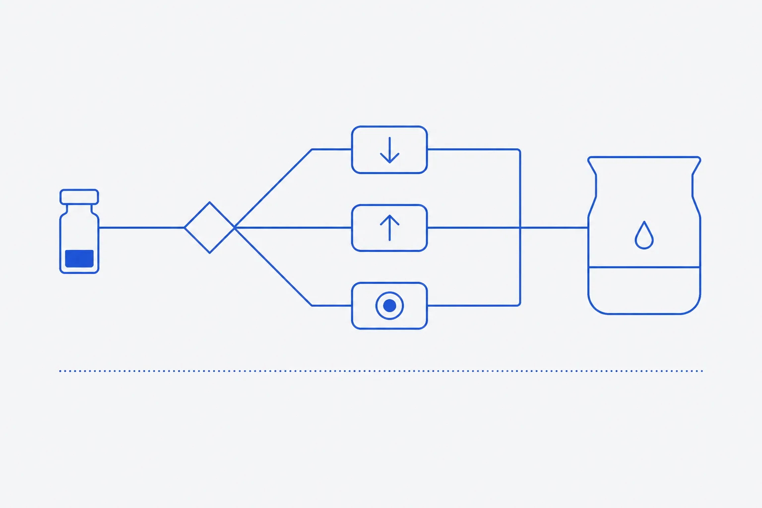
Solvent choice follows from the sequence: count the charged residues and let the result select the solvent — mildly acidic aqueous media for basic peptides, mildly basic media for acidic peptides, a small volume of organic co-solvent for near-neutral or hydrophobic sequences that resist water. Assay compatibility is the second filter, applied only to the solvents the chemistry already permits. Reversing that order — choosing what the assay likes and hoping the material dissolves in it — accounts for most dissolution failures.
Order of the decision
Two properties decide whether a sequence enters solution and in what. Net charge at the working pH is the first: a charged molecule is solvated by water and repelled from its own kind, and both effects keep it dispersed. Hydrophobicity is the second and works the other way — where non-polar residues dominate, the sequence associates with itself in preference to the solvent. Near-neutral and hydrophobic together is the difficult case, and the one aqueous media will not solve at any pH.
Assay compatibility is a real constraint but a second-order one. Every solvent below carries a downstream cost — an acid that shifts the pH of a small buffer volume, a volatile base that must be removed, an organic component that denatures protein reagents. Weigh those costs only among solvents that will dissolve the material. Selecting for assay convenience first gives the characteristic failure: a vial sat in buffer for an hour, cloudy, now harder to dissolve than it was dry, because partial dissolution followed by aggregation is not reversed by more buffer 2.
The order is fixed. Assess charge and hydrophobicity; identify the solvents that will dissolve the sequence; eliminate on assay grounds; then set the stock concentration so the final proportion of any problematic solvent falls below the assay's tolerance.
Net charge assessment
Net charge at neutral pH is estimated by counting ionisable groups. Effective pKa shifts with neighbouring residues, so the figure is approximate — but it is accurate enough to select a solvent, and it takes under a minute from the sequence.
- Count the basic residues: arginine, lysine, histidine. Add one for a free N-terminus.
- Count the acidic residues: aspartate, glutamate. Add one for a free C-terminus.
- Subtract acidic from basic. The difference is the approximate net charge at pH 7.
- Classify: +1 or greater is basic, −1 or lower is acidic, zero is near-neutral.
- Assess hydrophobicity separately, as the proportion of alanine, valine, leucine, isoleucine, methionine, phenylalanine, tryptophan and proline. A high proportion overrides the charge classification.
| Group | Site | At pH 7 | Note |
|---|---|---|---|
| Arginine | Guanidinium side chain | +1 | Charged across the practical pH range |
| Lysine | Amine side chain | +1 | Deprotonates only under strongly basic conditions |
| Histidine | Imidazole side chain | 0 to +1 | Titrates near neutral. Count as partial; basic in acidic media |
| Free N-terminus | Alpha-amino group | +1 | Absent if acetylated |
| Aspartate, glutamate | Carboxylate side chain | −1 each | Protonate under acidic conditions |
| Free C-terminus | Alpha-carboxyl group | −1 | Absent if amidated |
| Cysteine, tyrosine | Thiol, phenol | 0 | Ionise well above neutral pH; ignore here |
The rule follows from the count. A net positive peptide dissolves most readily in mildly acidic aqueous media, where the basic groups are fully protonated and the charge is largest. A net negative peptide dissolves in mildly basic media, the same reason inverted. At or near zero the peptide sits close to its isoelectric point in neutral water, with minimal charge and minimal repulsion between molecules, and is at its least soluble: move the pH away from that point, or introduce an organic co-solvent.
Hydrophobicity overrides the charge rule once high enough. A sequence dominated by non-polar residues resists aqueous media whatever its charge, and needs an organic co-solvent as the first addition rather than the fallback. Two failed aqueous attempts leave partially wetted, partially aggregated solid behind.
Solvent options
The table lists the solvents in ordinary laboratory use, the sequence type each suits, the proportion normally worked, and the compatibility cost carried downstream. Work down it unless the hydrophobicity assessment says otherwise.
| Solvent | Suits | Working proportion | Assay caveat |
|---|---|---|---|
| Sterile water | Clear net charge, low hydrophobic content. Always the first attempt | Neat | None of its own, but unbuffered: pH is set by the material's counterion and is not controlled |
| Dilute acetic acid | Basic sequences — net positive charge | 0.1% to 1% in water | Acidifies the receiving buffer. Volatile, so removable by lyophilisation. Confirm the buffer holds its pH |
| Ammonium bicarbonate or dilute ammonia | Acidic sequences — net negative charge | 10–100 mmol/L, or ammonia at comparable strength | Volatile and removable, but raises pH. Poor choice with cysteine present: disulfide exchange accelerates under basic conditions |
| DMSO | Hydrophobic and near-neutral sequences that resist aqueous media | Minimum volume, diluted to 0.1–1% final in most cell work | Oxidises cysteine and methionine; hygroscopic; biologically active in its own right. See below |
| Acetonitrile–water | Moderately hydrophobic sequences; material destined for chromatography | 10–50% acetonitrile — the lowest that works | Denatures protein reagents. Compatible with reversed-phase separation, where it is already the mobile phase |
| DMF | Strongly hydrophobic sequences where DMSO has failed | Minimum volume, diluted immediately | Poorly tolerated in most biological systems and harder to remove. Handle for its own toxicity |
DMSO is the general solvent of last resort, and it is not free. It oxidises cysteine and methionine over time — thiols to disulfides, methionine to the sulfoxide — so a stock changes composition across weeks even in cold storage, and oxidised species are aggregation-competent rather than simply lost 12. It is strongly hygroscopic: an opened bottle takes water from laboratory air, and a wet stock dissolves less well than anhydrous solvent. And it carries into the assay, where it is not an inert diluent but has its own effects on cells and protein reagents, at percentages easily reached from a concentrated stock.
The controls follow: anhydrous solvent from a small, recently opened container; the smallest primary volume the material allows; aliquots rather than one tube reopened repeatedly; and a solvent-only control at the identical final percentage in every experiment.
Dissolution procedure
Dissolve in the smallest volume of the strongest suitable solvent, then dilute into the working buffer — never the reverse. Buffer added to a solid that will not dissolve in it wets the surface, which hydrates and aggregates while the interior stays dry, and the material is then harder to dissolve than before the attempt 2. No further buffer recovers it.
- Equilibrate the sealed vial to ambient temperature before opening. Cold solid condenses water from laboratory air.
- Centrifuge briefly, or tap the vial down, so solid displaced onto the closure in transit returns to the base.
- Fix the target stock concentration and the final buffer volume before anything is added; together they set the solvent volume available.
- Add the minimum volume of the selected solvent down the inner wall, onto the solid rather than into the headspace.
- Allow the vial to stand undisturbed for several minutes. Time does more of the work than agitation.
- Agitate gently — slow inversion, or rolling the vial between the fingers. Do not vortex.
- Inspect against a dark background and then a light one, holding the vial still. Proceed only when clear of particles and haze.
- If solid remains, add one further small increment of the same solvent and repeat before considering a different one.
- Dilute by adding stock to buffer, slowly and with gentle mixing — not buffer to stock. Stock into the larger volume dilutes the peptide immediately; the reverse creates a local excess of buffer at the interface, where precipitation starts.
- Inspect again. Material coming out of solution on dilution has exceeded its solubility at the final composition; revise the stock concentration or the co-solvent proportion.
What not to do
Vortexing is the first avoidable loss. It generates shear and a large, continually renewed air–liquid interface. Peptides adsorb there, partially unfold, and desorb as aggregation-competent species; agitation is among the best-characterised mechanical drivers of aggregation 2. Foam is the visible marker. Gentle inversion mixes without the interface.
Prolonged sonication is the second. Cavitation applies extreme local mechanical stress, and a bath sonicator heats a small volume quickly — the operator does not notice, because the bath stays cool to the touch. Sonication therefore applies both aggregation-promoting stresses at once 2. Where it is genuinely needed, use short bursts with cooling between them, not as a first response to a slow vial.
Repeated heating is the third, and not a dissolution strategy. Warming may drive some fraction in, but temperature accelerates hydrolysis and deamidation, and thermally induced aggregation is generally irreversible — what comes out on cooling does not go back in 12. A sequence needing heat needs a different solvent. The same applies to standing in strong acid or base: those pH extremes are where the dissolved fraction degrades fastest 1.
Difficult sequences
Some sequences dissolve under none of the conditions above. Two classes account for most: those rich in hydrophobic residues, and those whose composition favours beta-sheet formation — runs of alanine, valine, isoleucine, leucine, phenylalanine, tyrosine, glycine and glutamine are typical. Such sequences self-associate into ordered, hydrogen-bonded structure that is thermodynamically stable rather than a kinetic accident 3.
Denaturing conditions will dissolve most of them. A chaotrope such as urea or a guanidinium salt, or a strongly hydrogen-bond-disrupting organic solvent, breaks the intermolecular structure, and the solid clears.
The consequence has to be stated plainly. Material that needed denaturing conditions is not stably dissolved; it is held apart by the denaturant. Dilution into aqueous buffer removes that condition, and the sequence is free to re-form the structure it was in before 23. What follows is visible precipitation, or — worse for interpretation — a clear solution of soluble oligomers, which are invisible, are not the monomer, and behave differently in any assay reading on concentration or activity.
Where this is anticipated: dilute slowly with mixing rather than in one addition, keep the final concentration as low as the assay tolerates, prepare immediately before use, and treat the result as conditional until an aggregation check has been run.
Filtration and clarity
Judge clarity after the solution has stood. A haze that clears on standing was air. A haze that persists — and a solution that scatters a beam held to its side — is undissolved solid or aggregate. It means an unknown fraction of the weighed material is not in solution, so every concentration calculated from that mass overstates what is present.
Filtration clears the appearance. A membrane filter removes the particulate and returns a visually clear liquid, and for many purposes that is the correct step. But the peptide in that particulate leaves with it, and soluble peptide is lost in addition by adsorption to the membrane, the syringe and the receiving vessel. Adsorptive loss depends on the surface material and is proportionally largest at low concentration 4.
So: if concentration matters, quantify before and after filtration by the same method and carry the post-filtration figure forward. Absorbance at 280 nm serves where the sequence contains tryptophan or tyrosine; otherwise use a colorimetric assay or quantitative chromatography against a standard. Filtering without quantifying converts a known problem — visible haze — into an unknown one: a clear solution of unrecorded concentration. Use low-binding labware, and keep it constant across a comparison 4.
Recording procedure
- Record the batch or lot reference against every solution prepared from it.
- Record the solvent by name and proportion: for a mixture, the composition; for an acid or base, its concentration.
- Record the mass taken, the solvent volume added, and the nominal concentration — stating whether it is calculated on gross weight or net peptide content.
- Record the dilution: volume of stock, the buffer it entered, the final volume, and the final percentage of any co-solvent.
- Record observations at each stage — time to dissolve, whether agitation was required, appearance before and after dilution, any haze or precipitate.
- Record any quantification, with method and both pre- and post-filtration values where filtered.
- Record the preparation date and the storage condition of the stock, and cross-reference it in every experiment drawing on it.
Recording the solvent matters because it does not stay in the background. It sets the pH, and pH is among the largest determinants of the rate at which dissolved peptide degrades 1. It sets the aggregation risk at dilution 2. And where it carries into the assay it contributes effects of its own, indistinguishable from effects of the peptide unless the solvent was recorded and a matched control run alongside. Two preparations of one batch in different solvents are two experiments, not one repeated.